Unseen Worlds
Revealing the science of healthier futures
Exhibition
Public
Art and science weave together in a new exhibition presented by SickKids Research Institute and OCAD University.
100 McCaul St.
, 2nd floor
100 McCaul St.
, 2nd floor
rgb(255,255,255)
Art and science weave together in a new exhibition presented by SickKids Research Institute and OCAD University.
Unseen Worlds is an immersive journey through hidden structures and stories shaping the future of children's health. It blends remarkable scientific images from SickKids labs with original artworks by OCAD U student and recent graduate artists, inviting curious minds of all ages to delve into the beauty, complexity and possibilities of scientific discovery.
From the smallest details inside our cells to breakthrough ideas that could transform care, Unseen Worlds shines a light on fascinating research and the promise of precision child health. The exhibition is guest curated by Golnoush Mir Salari.
The exhibition features imagery captured and created by more than 50 SickKids scientists across six research programs, and art that responds to this work by OCAD U student and recent graduate artists:
- TooPink
- Reymond Lise
- Jenna FitzGerald
- Sophia Kyungwon Kim
- Vivienne Valladares
- Sophie Dunkley
- Derek Berry
FAMILY DAY - SATURDAY, NOVEMBER 21
10 a.m. to 4 p.m.
Bring the whole family and discover science in a new way!
Explore incredible visuals from SickKids researchers and OCAD U, meet the scientists and artists behind them, and enjoy creative, hands-on activities designed for children and families.
Free and open to all ages.
rgb(255,255,255)
Anatomy of an Interface
Olga Sirbu, Post-doctoral Fellow
Rutka Lab
olga.sirbu@sickkids.ca
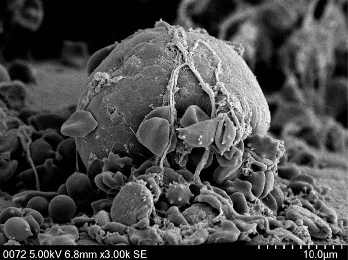
This image shows a single cell of medulloblastoma, the most common brain cancer in children, which has just landed on the surface of a spinal cord. We've captured it using a powerful "scanning electron microscope" that uses electrons instead of light to achieve an extreme zoom. Thin threads reach out, extending to hold the cell in place — a process called "leptomeningeal metastasis" that occurs when cancer cells spread through the fluid around the brain and spinal cord.
Right now, doctors have no way to stop leptomeningeal metastasis and, with no treatment available, children who develop it do not survive. That is why scientists at SickKids are studying how these cells survive in the fluid, searching for a weakness that could be exploited and usher in a new treatment to save children's lives
rgb(255,255,255)
Zebrafish - the heart(beat) of cardiac research
Esra Erkut, Graduate Student - PhD Candidate
Scott Lab
esra.erkut@mail.utoronto.ca or esra.erkut@sickkids.ca
One in 100 babies are born with congenital heart disease (CHD) and, for most, we don't know the cause. When researchers find a genetic change in a child with CHD, they can investigate whether it may be implicated.
Today, using the CRISPR gene-editing tool, we can create that same change in the matching gene of a zebrafish. Because zebrafish embryos are transparent, researchers can watch their hearts develop and beat in real time and, if an edited gene is indeed important for development, the zebrafish's heart may look malformed or have a weak pumping ability.
In this video we see, at left, a healthy zebrafish heart and, at right, one whose eif3s10 gene has been changed to model a specific SickKids patient with CHD. Its heart is thin and barely pumps blood, confirming that the gene mutation indeed caused the newborn's heart disease.
Finding the cause means that we can offer more personalized care while testing new treatments and building a deeper understanding of the developing heart.
rgb(255,255,255)
Interrupting the Invisible
Hui Guo
hguo@biochem.mpg.de
Gautier Courbon
gautier.courbon@gmail.com
John Rubinstein, Senior Scientist, Molecular Medicine
john.rubinstein@utoronto.ca
Here we present two videos detailing ATP synthase, a protein that acts as a tiny motor inside every cell. It sits in a cell membrane and uses the movement of protons to produce ATP, which stores and carries energy in cells. By studying how ATP synthase works, we gain a better understanding of one of the most basic processes of life — knowledge that can lead to new medications that can alter ATP production in harmful bacteria and cancer cells.
In the first animation, watch as ATP synthase rotates through its three main shapes.
The second reveals how the tuberculosis drug "bedaquiline" (shown in pink, blue and yellow) attaches to the protein so that it can no longer rotate and produce ATP, which leads to the bacteria cell's death. By visualizing these atomic structures, researchers can see exactly how the drug works and use that information to inform new treatment approaches to drug-resistant tuberculosis.
rgb(255,255,255)
The Immune Quiet Zone in Retinoblastoma
Helen Dimaras, Scientist
helen.dimaras@sickkids.ca
IMC was performed by Tina Chen, tina.chen@sickkids.ca
Retinoblastoma is a rare eye cancer in children and here we see the hidden cellular landscape of a child's eye as a healthy retina forms cancer. Like a map, it shows where different cells and proteins are located and how they are organised within the eye.
Each colour tells part of the story of the changing eye. Some colours show cancer cells growing quickly (blue). Others reveal the structure of the retina, while some highlight immune cells that help researchers understand how the body responds to disease (pink, red, green, cyan).
By studying these patterns, researchers can better understand retinoblastoma and develop more precise treatments for children, helping improve outcomes while reducing side effects.
rgb(255,255,255)
Celestial Paths of a Cellular Invader
Andrew Sydor, Research Associate
Brumell Lab
andrew.sydor@sickkids.ca
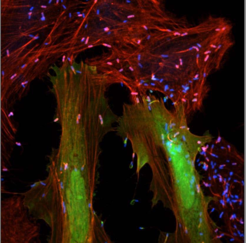
This image shows human cells infected with "Listeria monocytogenes", bacteria that can cause common issues like the flu and gastrointestinal problems and serious conditions including meningitis and sepsis. After entering a cell, Listeria take control of actin, a protein that helps cells keep their shape and use it to push themselves through the cell, leaving behind long trails called “comet tails.”
In this image, the bacteria are blue, actin is red, and a third protein is green. The researchers wanted to find out whether this green protein helps form the comet tails that allow the bacteria to move. By looking for places where the green and red signals overlap, researchers can learn more about how Listeria spreads inside and between cells. Overall, the work helps us better understand infection and identify new ways to prevent or treat bacterial disease.
rgb(255,255,255)
Strange New Worlds Within: The Architecture of Infection
Andrew Sydor, Research Associate
Brumell Lab
andrew.sydor@sickkids.ca
This model shows how Salmonella, a type of bacteria that can cause food poisoning, enters and survives inside human cells. The structures in this display are based on real protein models and have been enlarged millions of times so their shapes and connections can be seen.
At the centre is the Type III Secretion System (pink), a tiny needle-like structure the bacteria use to inject proteins into a cell. These proteins change how the cell works and help the bacteria create a safe place to live and multiply.
Scientists study the shape of these proteins to understand how Salmonella survives inside human cells. By seeing how bacteria interact with the cell, researchers can learn more about how infection develops and identify new ways to stop it.
rgb(255,255,255)
A Tumor's Tiny Twin
Nesrin Sabha, Research Project Manager
Rutka Lab
nesrin.sabha@sickkids.ca
James.rutka@sickkids.ca
saira.alli@unityhealth.to
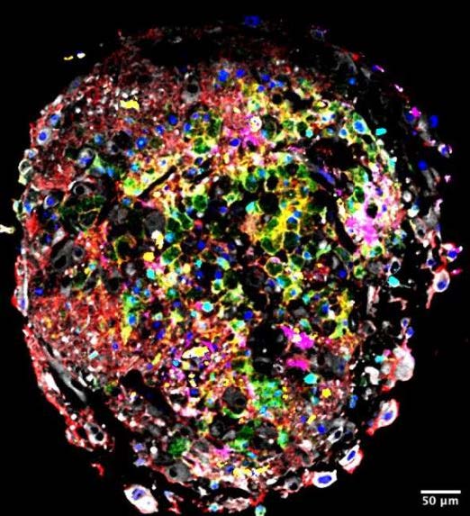
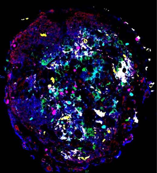
Here we see a microscopic, lab-grown version of a child’s brain tumour using their own cancer cells. Researchers wanted to know if these miniature tumours, called "organoids," could stay as complex as the original tumour. Each colour in the spectrum seen here marks a different type of cell, including tumour cells, immune cells and those that help build blood vessels.
Together, the colours reveal how that the organoid contains many of the same kinds of cells found in the patient’s tumour and shows evidence of how those cells interact. The image confirms that lab-grown models closely resemble the real tumour.
Using organoids, researchers can more closely study brain tumours in their search for new treatments.
rgb(255,255,255)
Guardians at Work
Nourhan Almasri, Graduate Student – MSc Candidate
Robinson Lab
nourhan.almasri@sickkids.ca
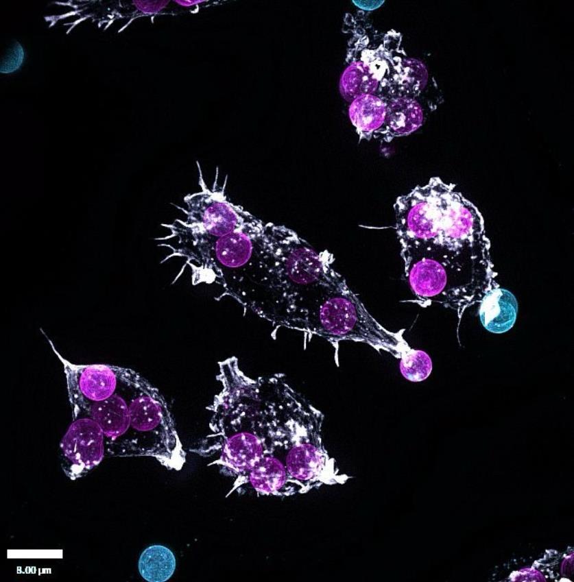
Here we find a team of macrophages (seen in white) in action, specialized immune cells that find, eat or ingest and destroy harmful invaders like bacteria, viruses, fungi, and parasites while removing damaged cells and cellular debris. The purple particles have been absorbed inside the macrophages while the blue particles remain outside.
By exploring how the immune cells take in and remove dangerous invaders, we can better understand why the immune system sometimes attacks the body's own healthy cells. This process can cause autoimmune disease and serious inflammation, for which new treatments are desperately needed. Doing so will also improve outcomes and care for children receiving life-saving organ transplants.
rgb(255,255,255)
Cells That Remember − Tracing Memory in the Hippocampus
Sung Mo Park, Research Associate
Josselyn Lab
sungmo.park@sickkids.ca
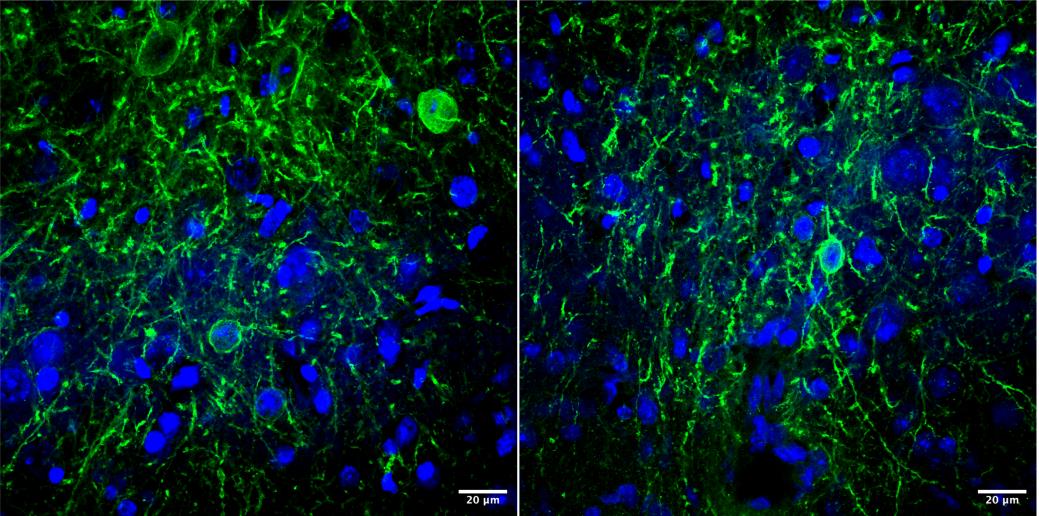
Here we find brain cells located in the hippocampus, the regione fo the brain that controls learning and memory. Researchers used a special tagging method to identify active cells in order to see possible "memory traces" in the brain — physical marks that experiences leave behind. The green are cells that illuminated while the brain learned something related to fear.
Memories exist in a complex group of brain cells that work together. By studying these activated cells, scientists hope to learn how memories form, last and are remembered later. This is especially important during childhood, when learning and emotions are prime aspects of a developing brain.
Unlocking the mysteries of memory may even help to explain brain development disorders or an assortment of related conditions.
rgb(255,255,255)
Shells
Jade Chan, Postdoctoral fellow
Xi Huang Lab
jsy.chan@mail.utoronto.ca
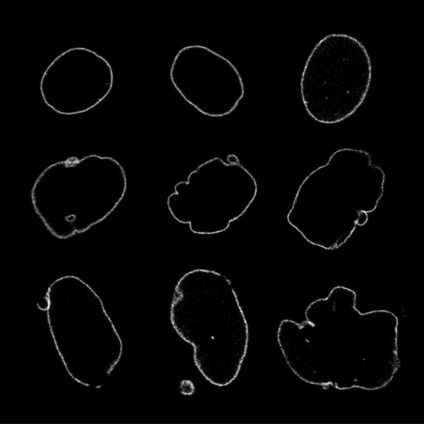
Medulloblastoma is the most common malignant brain cancer in children. Its cells grow rapidly and when they do, they squeeze tightly together putting pressure on each cell's nucleus where DNA is kept safe. (DNA carries instructions for making proteins, which build our cells and bodies.) Our lab found that a special protein called KCNG1 protects the nucleus from harm.
Here in this image we see two types of brain cancer cell nuclei. The top row shows cancer cells with KCNG1 which have smooth, solid nuclei that keep their DNA safe. The middle and bottom rows show cancer cells without KCNG1, which have nuclei that wrinkle, break apart, and leak damaged DNA.
This discovery, that KCNG1 acts as a protector, means that we have identified a new target to help treat this serious childhood cancer.
rgb(255,255,255)
Microglia: Finding Home in the Brain
Ai Tian, Postdoctoral Fellow
Muffat Lab
ai.tian@sickkids.ca
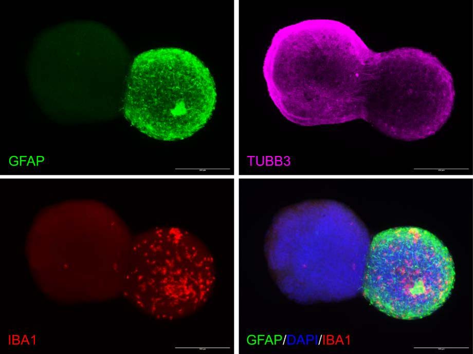
About one of every 10 cells in the brain are special immune cells called "microglia" that monitor the surroundings, keep things balanced and respond to germs or injuries. Unlike other major brain cells, microglia don't originate within the brain but instead migrate there before birth, as an embryo forms. They then establish lifelong residence in the brain, playing important roles in brain development, but we still don't know how wind up settling there.
Here in this image, we see microglia (right) interacting with an astrocyte (left). Researchers have used different fluorescent markers to help distinguish the cells and structures in order to [insert goal]. Describe what each is highlighting.
Our lab has found that when they move to the brain, microglia interact closely not with developing neurons but with astrocytes, which are supportive cells in the brain. This finding about early brain development opens new possibilities for cell-based therapies that could prove beneficial in treating neurological disorders.
rgb(255,255,255)
thank you, we love you.
Thomas Qu, Technologist I
Deshwar Lab
thomas.qu@sickkids.ca
Evelyn Popiel, Post-doctoral Fellow
Deshwar Lab
evelyn.popiel@sickkids.ca
Logan Newton, Graduate Student - PhD Candidate
Deshwar and Costain Labs
logan.newton@sickkids.ca
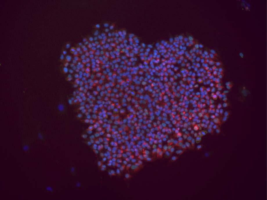
Here we witness a cluster of induced pluripotent stem cells (iPSCs) that were created from human skin cells. Using a method called electroporation, we added specific genes known as "Yamanaka Factors" into the skin cells. These genes allow the cells to switch identity and become stem cells. The blue and red colors show proteins that mark this new stem cell identity.
IPSCs are very valuable in research as they can become almost any cell type in the body. Our lab uses electroporation to reprogram skin cells from SickKids patients with genetic disorders, who have volunteered. We can then create cells related to each person's specific disease, such as neurons for brain disorders or liver cells for liver disease, which helps us understand how the disease works and test treatments using the patient's own genetic makeup. The ultimate goal is to use personalized, precision medicine to better understand and treat genetic diseases in children.
rgb(255,255,255)
The Inner Circle
Marynelle Icmat, Graduate Student - PhD Candidate
Brill Lab
marynelle.icmat@sickkids.ca
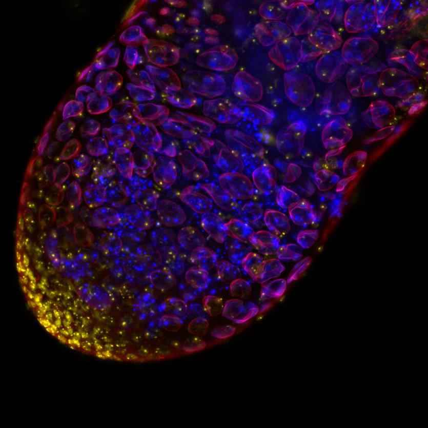
This image shows a testis from Drosophila melanogaster, better known as a fruit fly, with fluorescent stains used to highlight different parts of the tissue. Red shows LaminC, a protein that forms part of the nuclear envelope, the layer that Submission 16 surrounds and protects a cell’s nucleus. Blue shows proteins found close to LaminC, and yellow shows DNA.
The goal by our research team is to learn which proteins work with LaminC as sperm cells grow and mature. During this process, the nucleus changes shape and must stay organized properly, or it could lead to cells not functioning as they should. When the nuclear envelope does not form properly, it can affect fertility and growth.
In people, changes to Lamin proteins can cause diseases that affect muscles, the heart, and metabolism, often beginning in childhood. By studying how these proteins work together, researchers hope to better understand how these diseases develop.
rgb(255,255,255)
Lost in Translation
Brook Falk, Graduate Student – PhD Candidate
Brill Lab
brook.falk@mail.utoronto.ca
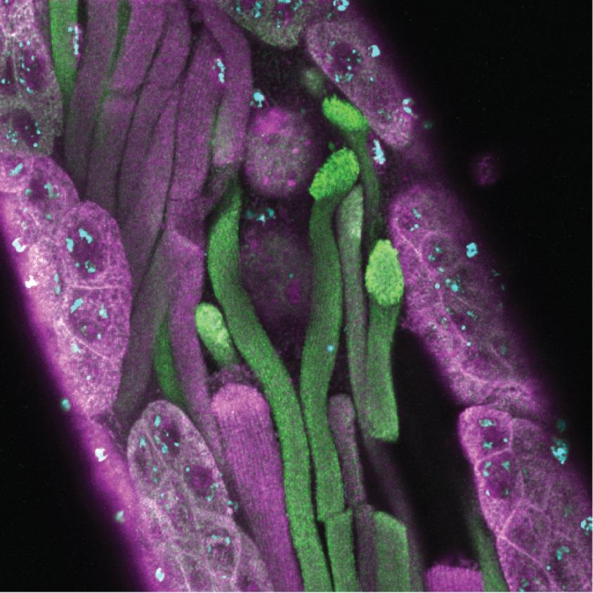
Here in this image, we see sperm cells growing inside the testis of a fruit fly. Two separate proteins, highlighted in green and purple, help control when and where other proteins are produced inside a cell. While the proteins are, our research team has found that they are present in different amounts as sperm cells grow — suggesting that they place distinct roles during sperm development.
Cells need the right balance of proteins to grow, divide and function properly. Imbalances create dysfunction and problems with proteins like these have been linked to diseases affecting the brain and nervous system, including cancer and neurodegenerative disorders.
By studying these proteins in fruit flies, researchers can learn how healthy cells work and what happens when these processes go wrong.
rgb(255,255,255)
Brain (in-a-dish) on Fire
Julien Muffat, Senior Scientist
Daniela Cobo, Graduate Student – MSc Candidate
Muffat Lab
daniela.cobo@sickkids.ca
julien.muffat@sickkids.ca
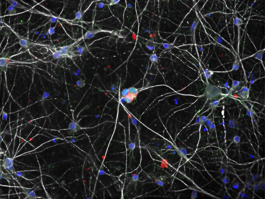
In this image we find the rare occurrence where a common childhood virus, HHV6, has infected human neurons that researchers have grown in a lab dish. Submission 18 White lines show the neurons' branching connections, similar to how they link up in real brains. Blue shapes are the nuclei, which hold the cells' genes. In the center, three nuclei show extra colors. Green specks reveal the virus has infected these cells and started making its own proteins while red staining reveals the virus has moved further into its life cycle. Scientists now wonder: will it damage these cells, spread further, or be destroyed by the immune system?
The HHV6 virus typically causes only a mild rash and fever in children, but in rare cases it can lead to serious brain complications — and because it remains in the body for life, evidence suggests it may play a role in later diseases such as Alzheimer’s disease or multiple sclerosis. Researchers are studying what makes certain brain cells and children more vulnerable to this outcome for whom these lab-grown cells could help predict disease risk and guide personalized treatments.
rgb(255,255,255)
Signalling constellations
Elissa Goodbrand, Graduate Student – PhD Candidate
McGlade Lab, In collaboration with the SickKids Imaging Facility
elissa.goodbrand@sickkids.ca
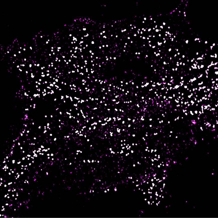
At first glance, this image resembles a map of constellations but in fact this reveals a hidden world inside a human cell. The bright spots are clusters of a protein called Numb, which helps cells organize and send messages.
Researchers have used a powerful microscope to see details that are too small to be viewed with a regular microscope. Here, they’ve discovered that Numb is not spread evenly throughout the cell but instead it gathers into small glowing bunches. It’s important to understand how Numb clusters and what they do inside cells because certain forms are found at higher levels in several childhood cancers. By understanding how Numb is organized, scientists hope to better understand the biological changes linked to childhood cancer
rgb(255,255,255)
Spots and Stripes
Nigel W. Griffiths, Lab Research Project Coordinator
Brill Lab
nigel.griffiths@sickkids.ca
Zebrafish, which at full growth are about 5 cm long, are well-used in medical research. This is because we know a great deal about their genes and development (roughly 70% of their genes are shared by humans) and offer unique biological and practical advantages for lab studies.
They are particularly beneficial when we study early development, as females can lay hundreds of eggs at once, which grow outside her body and are transparent meaning we can use microscopes to watch organ development, cell division and disease progression in real time. In just a few days, a single fertilized egg grows into a baby fish with a beating heart, working organs, and a fish-shaped body.
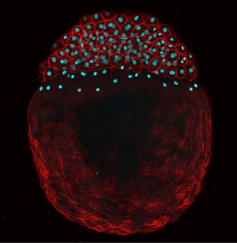
This image shows a zebrafish embryo preserved three hours after fertilization, stained in cyan to reveal DNA and in red by show actin, which outlines cell shape. At this stage, the embryo looks like a small ball of cells attached to a large yolk that provides nutrients for growth. In each cell, a nucleus is home to DNA and by studying which genes are active early on, researchers can learn how single cells become complex bodies.
rgb(255,255,255)
Airway epithelium
Dr. Kai Du, Research Associate
kai.du@sickkids.ca
Dr. Tarini Gunawardena, Laboratory Research Project Manager
tarini.gunawardena@sickkids.ca
Dr. Christine E Bear, Senior Scientist
bear@sickkids.ca
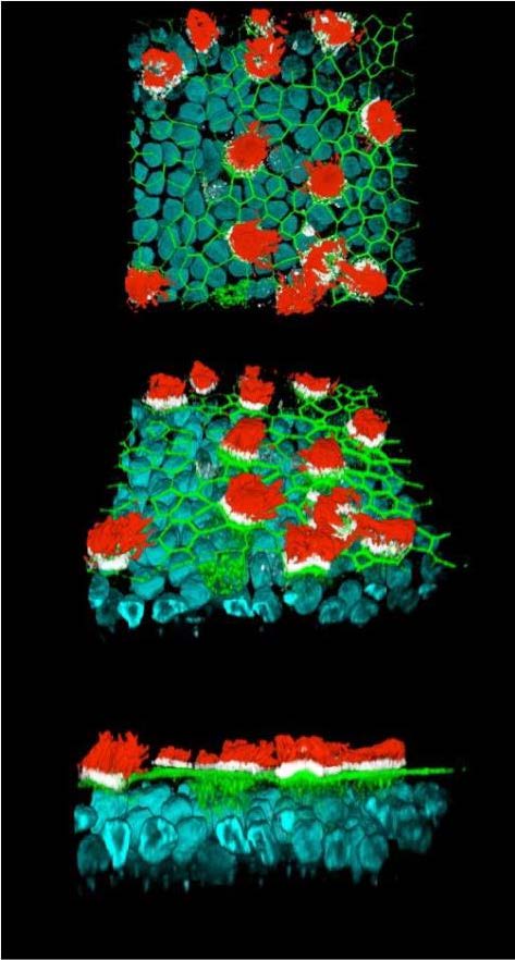
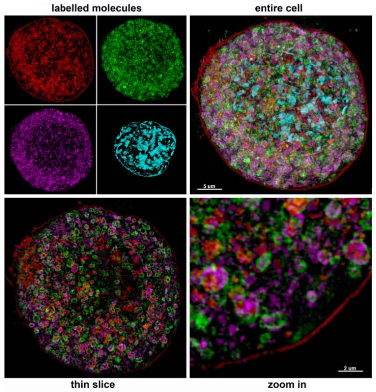
In this image we find a healthy human airway epithelium — the layer of cells that lines the airways and helps protect the lungs. Blue marks the nuclei, which contain each cell’s genetic material. Green shows where neighbouring cells connect to form a protective barrier. Red reveals cilia, tiny hair-like structures that sweep mucus, dust, and germs out of the airways. White marks the surface of the cells where the cilia are attached.
Cystic fibrosis is an inherited disorder (whose genetic cause was famously found at SickKids) that primarily affects the lungs. Researchers are analyzing these images to understand why cystic fibrosis causes thick mucus to build up in the lungs, which causes frequent infections. By comparing tissues from people with and without cystic fibrosis, they can see how changes in the lining of the airways impact lung health.
rgb(255,255,255)
The Architecture of Vision
Mercedesz Praszner and James Li, Graduate Students – PhD Candidates
Lefebvre Lab
mercedesz.praszner@mail.utoronto.ca
james.li@sickkids.ca
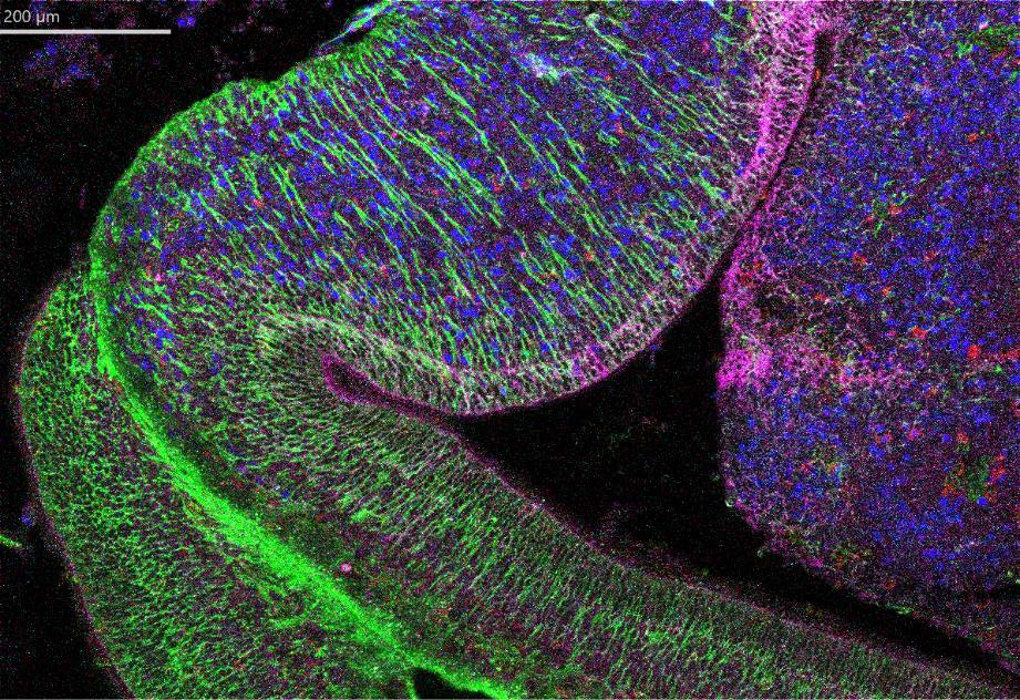
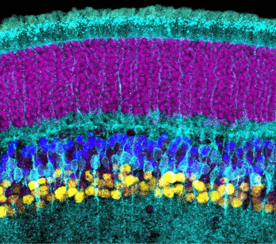
This image shows a slice of a developing retina, revealing how its cells are neatly arranged to support vision. The top layer (in magenta) holds photoreceptors, which sense light and initiate visual signals. These signals travel through inner circuits made of bipolar cells (blue) and amacrine cells (yellow), then reach ganglion cells, which send visual information to the brain. Müller glia (cyan) are like the mechanical scaffolding, stretching across the whole tissue, providing support and helping keep the retina organized as it grows. As the majority of research on retinal disease focuses on photoreceptors, less is known about how the inner circuits form and stay functional—where problems can begin to compromise vision. Good vision depends on these circuits forming correctly early on, when some cells die off, so the right mix of cell types remains.
By studying how genes control cell survival and circuit-building, this research helps explain how vision networks form. It could lead to better understanding of retinal disorders and new treatments for children with vision problems.
rgb(255,255,255)
Structures of Cerebellar Neurons
Benjamin-Israel Moke, Graduate Student - PhD candidate
Lefebvre Lab
benjamin-israel.moke@sickkids.ca
James Li, Graduate Student - PhD candidate
Lefebvre Lab
james.li@sickkids.ca
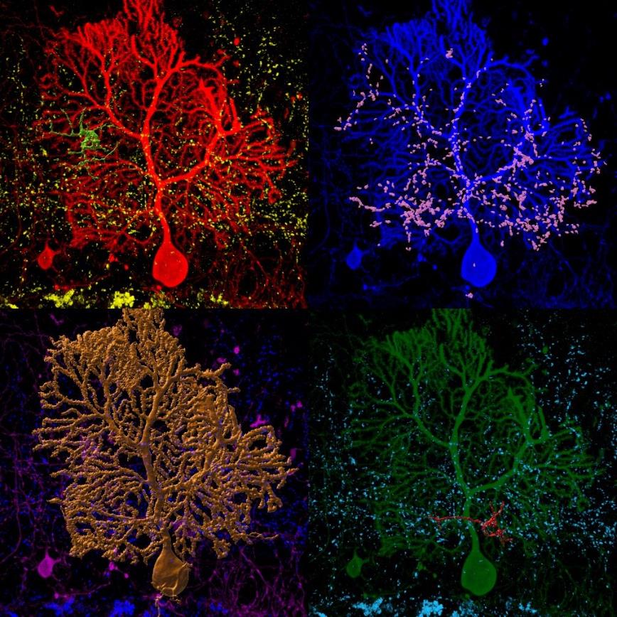
The brain has many types of neurons that use electrical impulses and chemical signals to transmit and coordinate information across the entire body. Submission 24 The Purkinje cell is a large neuron in the cerebellum that connects with many nearby cells, helping to coordinate movement and balance. Like in most regions of the brain, cerebellum cells are tightly tangled together, but researchers can use 3D fluorescent microscopy to isolate Purkinje cells from their neighbours.
In this image we see a Purkinje cell (gold) alongside connecting cells in different colors: a green cell and a red cell link to different parts of it, while pink marks many smaller connections coming from elsewhere in the brain. By building 3D models of these neurons, scientists can spot when connections fail to form correctly and thereby study how brain connections change in developmental disorders.
rgb(255,255,255)
At the Crossroads of Repair and Connection
Lisa Gazdzinski, Research Associate
Wheeler Lab
lisa.gazdzinski@sickkids.ca
Benjamin-Israel Moke, Graduate Student - PhD candidate
Lefebvre Lab
benjamin-israel.moke@sickkids.ca
Xi Zhou, Graduate Student - PhD Candidate
Wheeler Lab
xi.zhou@sickkids.ca
Larry Afun, previous undergraduate project student in Anne Wheeler’s lab, now MSc candidate in Veronique Miron’s lab at St Michael’s Hospital
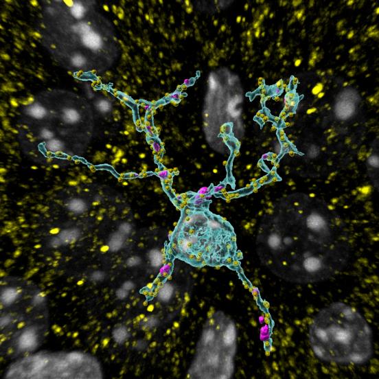
Concussions are common among children, and in about one-third of cases, they experience cognitive and emotional symptoms that linger for years afterward. In the Wheeler Lab at SickKids, we study what happens to brain cells that might cause these symptoms, so we can understand the injured brain and design better treatments.
Microglia are specialized immune cells in the brain and its first line of defense, scanning their surroundings for infections, toxins and cellular damage, and then responding as needed. This image shows a microglia cell that tests positive to a marker CD68 (magenta), which means it is clearing away cellular debris to help repair tissue.
Microglia also shape the brain's wiring by trimming synapses — the connections between nerve cells. The yellow dots are excitatory synapses, letting us see how microglia and synapses interact in an injured brain. This research offers insight into what causes concussion symptoms and points toward possible treatments.
rgb(255,255,255)
Starburst cells are the motion detectors in our retina
Samantha Ing-Esteves, former PhD Student
Lefebvre Lab
Julie L. Lefebvre PhD, Senior Scientist
Julie.lefebvre@sickkids.ca
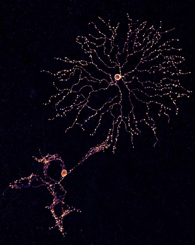
At the back of the eye sits the retina, a thin layer of nerve tissue that senses light and sends visual information to the brain. It contains more than 100 types of nerve cells, each with its own shape and job.
These microscopy images reveal "starburst" neurons, named for their firework-like branches, which are essential for vision because they detect motion and direction. Here our research team uses fluorescent labels to show two starburst cells in detail.
At top right, the cell reveals a normally developing retina — its branches spread out evenly, giving it plenty of room to form connections in the outer ring of the eye. At bottom left, the cell's branches don't spread apart but instead overlap and tangle together, a pattern that weakens its ability to detect motion. We can see that it is missing specific proteins, called Protocadherins.
By showing how these neurons wire themselves correctly, the research helps explain how children's visual systems develop the ability to track moving objects and keep images steady while they move.
rgb(255,255,255)
Pulling Tubes from Fiery Donuts
Ashwin Ian D’Souza, Post-doctoral Fellow
Brill Lab
ashwin.dsouza@sickkids.ca
Paul Paroutis, Service Manager Research Core Facility (Imaging)
paul.paroutis@sickkids.ca
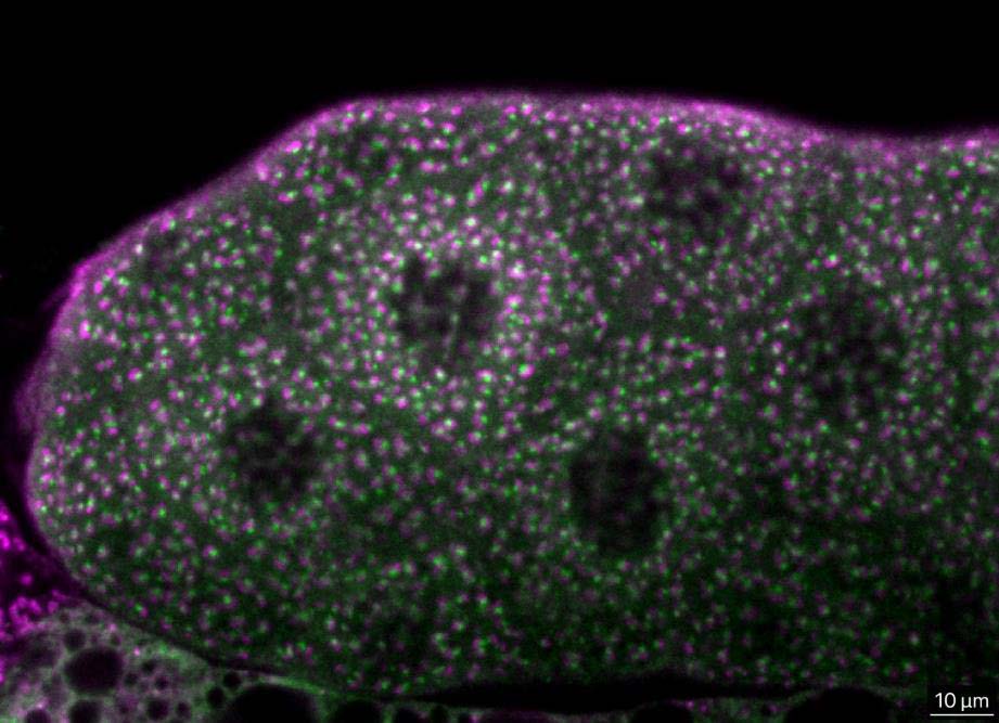
In this video, watch tiny structures move inside living cells from the salivary glands of fruit fly larvae. Researchers have used fluorescent tags, which illuminate parts of cells, so they could follow structures called PI4KII tubules.
These tubules stretch out from round cell compartments called endosomes, making them look like bright, glowing donuts. The tubules travel along microtubules, which act like tracks inside the cell. Researchers are studying these tubules to learn what they do and where they go in a bid to understand how tubules move materials around the cell.
This is important because cells need a constant flow of materials to work properly and by learning how they move from place to place, we can better understand how cells stay healthy and what happens when such cellular transport is dysfunctional. This may help researchers learn more about diseases that affect children.
rgb(255,255,255)
Big Brain Energy
Florence Chu, Graduate Student - MSc Candidate
Brill Lab
florence.chu@sickkids.ca
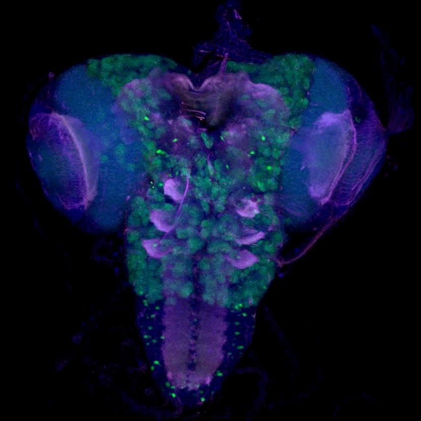
Here in this image we find the brain of a young fruit fly, often used in research because parts of their brain development are similar to those in humans.
In green we identify brain stem cells, which are special cells capable of making new ones. In blue is the nuclei, which is home to each cell's genetic instructions. Pink forms the outer edges of cells. This is part of ongoing research into how stem cells divide and build a developing brain — a process of vast importance and tremendous growth before birth and during early life. When stem cells do not divide properly, the brain can grow much smaller than expected, a condition called microcephaly.
By adding human genetic variants to fruit flies and studying their brains, researchers can learn which genes are important for healthy brain development in children.
rgb(255,255,255)
The seeds of life from nebula to comet (a series)
Lala Fabian
Brill lab
lacramioara.fabian@sickkids.ca
This image shows a male reproductive cell from a crane fly insect at the very end of cell division. At this stage, one cell is splitting into two new cells, a vital process that helps living things grow, develop, and produce new cells.
To divide successfully, the cell must reorganize its internal support system, called the cytoskeleton, which ensures the cell preserves its shape and ability to move its parts. During cell division, some of these structures gather in the middle of the cell to form a ring. This ring slowly tightens, like pulling a drawstring on a bag, causing the cell to pinch inward until it separates into two cells. Inside the cytoskeleton are protein strands called actin filaments, which create the force needed for the ring to tighten and complete the division process.
Here scientists have made the actin filaments visible using a special fluorescent dye that attaches to them glows brightly. The bright ring in the center of the image reveals actin working to divide the cell into two. This research technique helps scientists better understand how cells divide and how living organisms grow and reproduce.
rgb(255,255,255)
A cell full of surprises
Fred Pluthero, Lab Manager
Kahr Lab
fred.pluthero@sickkids.ca
Helen H. Y. Yao, Graduate Student - PhD Candidate
Kahr Lab
helen.yao@sickkids.ca
Walter Kahr, Principal Investigator
walter.kahr@sickkids.ca
Here we have a video of a megakaryocyte, a large cell in the bone marrow that makes platelets — tiny cell fragments in the blood that stick together to form clots and stop bleeding. When they don't work properly, the body has a harder time stopping bleeding and it can lead to certain disorders.
Researchers are studying how megakaryocytes make platelets and the small packets inside them that carry important materials. The spectrum of colours here mark different parts of the cell, including its genetic material, outer covering, and a protein called von Willebrand factor (VWF), which helps blood clot.
The video moves through thin slices of the cell so researchers can look inside and see these packets more clearly. By studying how Submission 31 megakaryocytes make platelets, researchers can better understand typical development as well as find cellular clues about why some children experience bleeding problems.
rgb(255,255,255)
Zebrafish models of adolescent idiopathic scoliosis
Dr. Brian Ciruna, Senior Scientist and Director of the Developmental, Stem Cell & Cancer Biology Program
ciruna@sickkids.ca
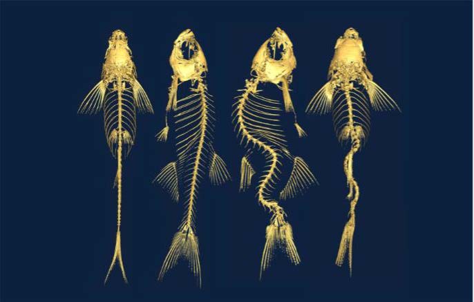
Adolescent idiopathic scoliosis (AIS) is a spinal deformity that usually shows up during puberty in otherwise healthy kids. It affects about 4% of children worldwide, and severe cases can cause chronic back pain and disability. Scoliosis was first described by Hippocrates, making it one of the oldest known birth defects. Even so, its causes remain unclear, and treatment is still limited to long-term bracing or major surgery.
Progress has been slow, partly because we couldn't study it in preclinical models. Humans walk upright and our spines compress from head to toe, and the curves of scoliosis are a uniquely human condition.
Nonetheless, fish experience spinal forces much like ours as they swim forward through water. SickKids researchers created the first genetically engineered zebrafish models of idiopathic scoliosis. These fish develop rotating spinal curves during growth that closely resemble the human disease, offering new clues about its causes and possible treatments
rgb(255,255,255)
Between Sequence and Self
Jocelyn Lee, Project Coordinator
IP&C, Led by Stephen Scherer, Chief of Research, SickKids
Jocelyn.lee@sickkids.ca
In this piece of art, we demonstrate how small changes in our DNA can affect how we experience the world.
It's based on SickKids research on PTCHD1-AS, a gene linked to key traits of autism spectrum disorder (ASD). The piece follows a path from gene to brain to behavior. An X chromosome expands into a brain image, with colored sections representing different behaviors. These sections reflect a major finding: changes in PTCHD1-AS relate to differences in social interaction and repetitive behaviors, while learning and memory stay unaffected. In lab studies, disrupting this gene increased repetitive actions and reduced social interaction, without harming broader thinking skills.
The artwork resembles a scientific poster or lab notebook, mixing digital design with hand-drawn pencil work. It creates tension between scientific precision and human complexity, showing that even as we learn more about ASD's biology, each person's experience remains unique.
By linking molecular changes to behavior through art, this piece reflects SickKids' work toward understanding neurodevelopment, while honoring the nuance science still has to explore.
rgb(255,255,255)
Protecting the gift Didactic
Ori Scott, Immunologist and Scientist
Ori.scott@sickkids.ca
Spencer Freeman, Senior Scientist
Spencer.freeman@sickkids.ca
Blayne Sayed, Surgeon and Associate Scientist
Blayne.sayed@sickkids.ca
Benjamin Steinberg, Anesthesiologist and Scientist
Benjamin.steinberg@sickkids.ca
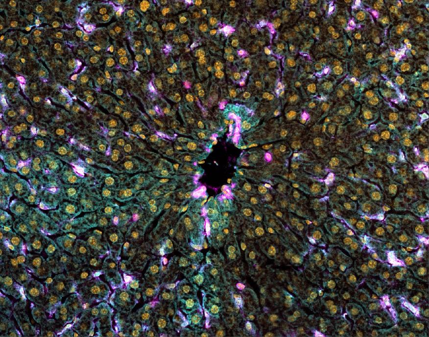
This image shows a human liver. Different colours highlight different parts of the tissue, including showing immune cells (magenta), architecture (cyan), and cellularity (yellow).
The research team is investigating what happens at a cellular level during a liver transplant in an attempt to protect donor organs from damage that can occur. That's because, when a donor liver from one person to another, blood flow must be stopped for a short time — and when it returns, it runs the risk of what's called ischemia-reperfusion injury. Submission 34 This fall, 40 years since the first liver transplant performed at SickKids, our researchers made a breakthrough discovery: they found that a protein called NINJ1 plays a previously unknown and important role in this process. NINJ1 can cause dying cells to break open and release substances that harm nearby tissue. In their study, blocking NINJ1 reduced liver damage in pre-clinical models. The team hopes this research could lead to new ways to protect donor livers before transplantation, which could help improve liver transplant outcomes for children and adults who need a new liver.
rgb(255,255,255)
Tics
Grace Leo
Elia Lab
Tourette syndrome (TS) is a neurological disorder that may cause sudden unwanted and uncontrolled rapid and repeated movements or vocal sounds called tics. This piece, done with watercolours and permanent marker, highlights the impact of TS on patients, aiming to bridge the gap between data from thousands of people with the actual experience of living with it. It is derived from an original figure that went with a review of studies involving 1,331 patients — depicted here by 1,331 water drops. Each drop is unique, representing one person who has taken time to share their experience of TS with researchers to help it improve our understanding of the condition. The tics people experience are the result of an irresistible urge, similar to a scratching an urgent itch. The artist/researcher has lived with TS since age four, and describes this urge as a constant dripping on their forehead. Hence this analogy of the ripples from a water drop, showing how tics impact a person's own psyche and how they interact with the world.

rgb(0,0,0)
rgb(0,0,0)
TooPink
TooPink is an interdisciplinary emerging artist based in Toronto and a student at the Ontario College of Art and Design University. Her work spans many mediums including film, performance, sculpture, installation, and analogue and experimental photography. She finds inspiration through history, identity, representation, and family, often exploring ephemerality and materiality in her work. She works with historical photographic processes, studying methods first recorded in the 1800s and experiments with how historical techniques can situate a modern artistic practice.
Artwork Statement
Study of Body, cyanotype, 2025.
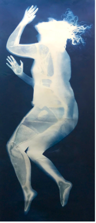
Study of Body is a life-size print of a human body created using a photographic technique invented in the 1800s. The work explores the history of photography in scientific studies, citing cyanotype’s use in creating the first photographically illustrated book, a study of botanical specimen. The print was created by placing the body onto a photo-sensitive surface and exposing to sunlight, creating a perfect imprint of the body reminiscent of x-ray prints.
rgb(0,0,0)
Reymond Lise
Reymond Lise is a Toronto-based interdisciplinary queer artist with a deep appreciation for nature. His art comes from a desire to interact with the world through compassionate scientific curiosity. He gives attention to the ephemeral by exploring seemingly simple subjects that, upon closer inspection, reveal new worlds. Using an integrative approach to problem-solving, Lise synthesizes artistic and scientific methodologies into a multifaceted practice that marries both fields in a uniquely engaging way. Through his art, he invites others to appreciate the wondrous complexities that emerge when life is viewed through an inquisitive lens.
Artwork Statement
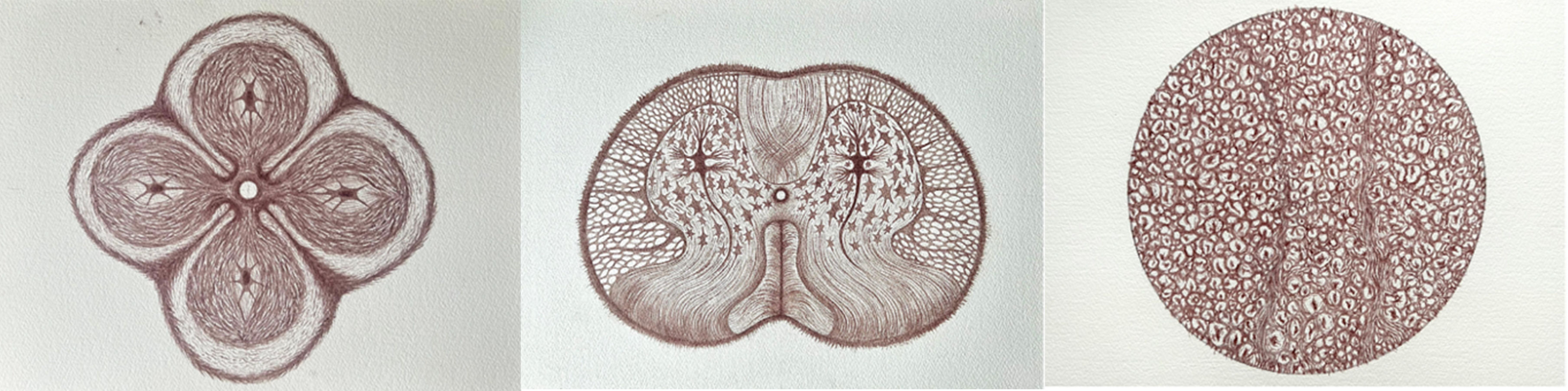
Fading Sensations, Fading Movements, Fading Memory explores the intricacies of neural structures through meticulous illustration of spinal cord tissue using homemade, non-archival ink. The mind, as we conceptualize it, is no tangible thing. There exists the physical structure: the brain, spinal cord, nerve fibers, but where in this system does the mind reside? Our actions, thoughts, and memories emerge from the firing of specific neurons in specific sequential patterns. They are the collective action of 100 billion cells sharing 100 trillion connections. From this enchanted loom, our self emerges. As we age, this current slows, connections fray, and some are lost altogether. The impermanence of the ink speaks to this ephemerality. It is daunting, but inevitable. All beautiful things must come to an end.
rgb(0,0,0)
rgb(0,0,0)
Jenna FitzGerald
Jenna FitzGerald is a multidisciplinary artist from Toronto whose work explores the intersection of technology and nature, drawing on both organic and digital imagery and materials.
Her artworks explore impermanence in the digital age and create new mythologies to reveal the magic in the world all around us, as well as themes that reflect our current moment of existence within the Anthropocene epoch. FitzGerald employs a variety of mediums throughout her work, including photography, film, graphic design, 3D modelling, and experiential installations.
Artwork Statement
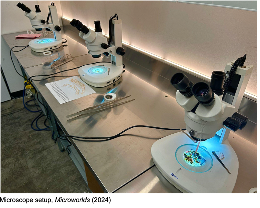
Microworlds of Absurdity is an interactive installation that invites viewers to create collages using images that have been drastically miniaturized—small enough to be explored only through a microscope! The premise behind the work is that regular-sized artworks in the world have suddenly been shrunk to a minuscule scale, allowing us, as scientists, to examine them through microscopes. The slides featured in this installation include a variety of images of iconic artworks, allowing visitors to playfully engage with art history by combining the disciplines of art and science together.
rgb(0,0,0)
Sophia Kyungwon Kim
Sophia Kyungwon Kim is a Toronto-based visual artist whose multidisciplinary practice materializes the invisible labor of domestic and parental care. Drawing from a 22-year career in public safety and the lived experience of raising four children, her work investigates care as a durational enactment of responsibility and vital social infrastructure. Holding an MFA from OCAD University, Kim seamlessly bridges traditional and digital methodologies—moving from oil painting and hand-sewn textile installations to 3D scanning and bronze casting. Her practice reclaims institutional and domestic maintenance as an enduring, generative force that sustains life.
Artwork Statement
Title: Weaving the Unseen
Medium: Fabric, stuffing, thread, and bronze
Size: Approx. 2m x 5m (Dimensions Variable)
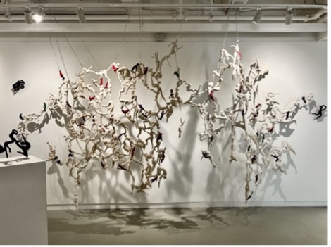
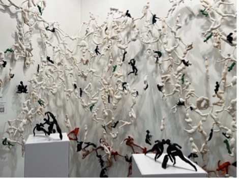
Weaving the Unseen is a monumental, site-specific installation that bridges the intimate devotion of daily caregiving with the essential, supportive networks of ecological mycelium. Spanning 2m x 5m, the installation integrates heavy bronze modular elements with an expansive wall-mounted matrix of hand-sewn textile figures, each meticulously crafted to capture the emotional moments and physical gestures of everyday care.
The figures interconnect through "anastomosis"—a biological structural joining—mirroring the networks of human care and natural interdependence. By transmuting soft, vulnerable fabric forms into permanent bronze components via 3D scanning, the work materializes the profound physical and emotional resilience inherent in recovery and caregiving. Ultimately, this immersive installation frames "maintenance" not as an invisible burden, but as an active, vital matrix that quietly sustains and protects our collective life.
rgb(0,0,0)
Vivienne Valladares
Vivienne Valladares is a Toronto-based interdisciplinary artist working across animation, immersive installation, drawing, painting, textile and sculptural work. She holds an MFA in Interdisciplinary Art, Media and Design from OCAD University (2026) and a BFA in Digital Media Arts from Srishti Institute of Art, Design and Technology (2022). Her practice is rooted in storytelling with tenderness and wonder, exploring both the intersections of art and science and the quiet, intimate labour of care and love. Recent exhibitions include her MFA solo show All the Light Reveals (2026) and Love Letters (2025), which she curated and exhibited in.
Artwork Statement
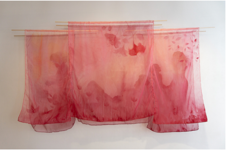
love = duty is a textile work honouring the quiet, laborious and oftentimes dutiful acts of caregiving. Created with acrylic paint and hand-embroidery on translucent organza, it takes its title from words my father spoke during my mother's illness. In one of our many conversations about the caregiving my mother required, he said, “love and duty are both four-letter words and mean the same thing to me.”
In this piece, I depict tender moments of care I have been fortunate to witness and receive — the small, repeated gestures that carry the weight of love. Painting, embroidering, and stitching delicate fabric became their own labour of love, mirroring the patience and endurance that caregiving demands. The semi-transparency of the fabric is much like memory itself, oftentimes obscured and layered. love = duty speaks to the resilience and strength behind oncological and palliative care. It pays homage to the unseen hours of devotion that sustain healing long before or after it is visible.
rgb(0,0,0)
Sophie Dunkley
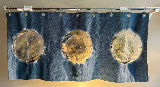
Petri, 2024
Alginate bio-plastic, artificial sinew, fabric scraps, assorted shells and tumbled rocks from the shores of my hometown
Petri explores the relationship between macro and micro ways of seeing through a lens-based approach to observation. Created using alginate bioplastic, the work translates organic forms into an active site that shifts between the familiar and the microscopic. Viewed from a distance, Petri resembles a contained object or landscape; upon closer observation, the forms begin to parallel our own cellular structures, hinting at a biological world that might otherwise remain unseen...
I invite you to move closer, look differently, and reconsider what you are seeing.
The curtain that partially conceals the work, functions as a membrane between the visible and invisible. Pulling it back becomes an act of observation, it is to reveal a space that feels simultaneously intimate yet unfamiliar. Through the use of alginate bioplastic and fabric scraps, Petri considers how artistic processes and experimental media can echo scientific methods of looking; transforming observation into an embodied and material experience.
rgb(0,0,0)
Derek Berry
Derek Berry is a multidisciplinary artist based in Orangeville, Ontario who works in varying printmaking mediums alongside installation. He specializes in intaglio, relief and serigraphy printmaking techniques. Currently Derek is continuing his undergraduate studies at the Ontario College of Art and Design University (OCADU) majoring in printmaking and publications and holds a 3-year diploma from Georgian College for fine arts. Derek has exhibited predominantly in the greater Toronto area and southern Ontario, has co-founded the PRNT Collective in Barrie, and has previously been awarded an emerging artist’s residency in Atlanta, Georgia.
Artwork Statement
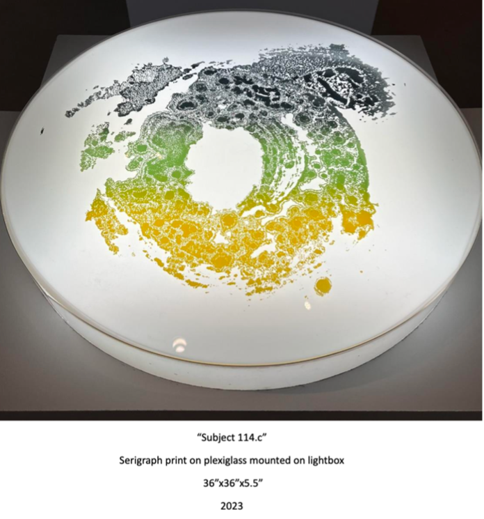
'Subject 114.c' is a part of a series of installations titled 'Macro-Organisms', whose purpose is to emulate and exaggerate the unseen contaminates that cause myself immense distress when navigating the outside world. This grief is caused by obsessive-compulsive disorder (OCD), a mental disorder whose unrelenting intrusive thoughts cause intense fear and debilitation towards the focus of one's obsession. During and following the Covid-19 pandemic my disorder solely focused on pathogens, and it is this heightened awareness towards the unseen that I have made tangible and visually perceivable within these subjects. It is my hope that the shared experiences of uncertainty, isolation, and fear caused by the recent pandemic in relation to these larger-than-life organisms permits the viewer to further their own understanding of obsessive-compulsive disorder.
rgb(255,255,255)
rgb(255,255,255)

rgb(255,255,255)

Please be advised that OCAD U hosted events may be documented through photographs and video. These images may be used by the University for promotional, advertising, and educational purposes. By participating in our events, both on campus and off-site, you consent to allowing OCAD University to document and use your image and likeness. However, if you do not want us to use a photo or video of you or your child, please don’t hesitate to let us know when you arrive at the event. You’re also welcome to get in touch with OCAD University’s Marketing & Communications office: communications@ocadu.ca.
Be mindful of those in our community who have scent sensitivities; please help OCAD U maintain a healthy, scent-free campus.